A Beginner’s Guide to ATAC-seq
Introduction to ATAC-seq
Gene regulation is fundamental to understanding cellular function and adaptability. A crucial aspect of this regulation is chromatin accessibility, which refers to the openness of various genome regions to transcriptional machinery. The ATAC-seq (Assay for Transposase-Accessible Chromatin using sequencing) technique is a groundbreaking method designed to map chromatin accessibility across the entire genome. It provides a detailed view of which regions of the genome are open and potentially active, offering valuable insights into gene regulation and epigenetic modifications.
ATAC-seq presents several advantages over older techniques such as DNase-seq and MNase-seq. It requires fewer cells, produces faster results, and offers a more nuanced view of the chromatin landscape. These benefits make ATAC-seq especially useful for studying complex biological systems and rare cell types.
Understanding Chromatin Accessibility
To grasp the significance of ATAC-seq, it’s essential to understand a few foundational concepts about chromatin structure and function. Chromatin is composed of DNA and histone proteins that package the DNA into a compact, organized structure. This packaging can either be open (euchromatin) or closed (heterochromatin). Open chromatin regions are more accessible to transcription factors and regulatory proteins, whereas closed regions are less accessible.
The positioning of nucleosomes, the fundamental units of chromatin consisting of DNA wrapped around histone proteins, plays a critical role in chromatin accessibility. Regions of DNA that are free from nucleosomes are often more accessible and associated with active transcription. Additionally, open chromatin regions frequently contain binding sites for transcription factors, which are crucial for regulating gene expression.
The ATAC-seq Workflow
The workflow of ATAC-seq involves several key steps, each contributing to the generation and analysis of chromatin accessibility data:
- Sample Preparation: The first step in ATAC-seq is isolating a small number of cells while preserving their chromatin structure. Common sources for ATAC-seq samples include cell lines, tissue samples, or primary cells. It’s crucial to minimize contamination and maintain cell viability to obtain reliable results. Proper sample preparation ensures that the chromatin remains intact and representative of the biological state being studied.
- Transposase Reaction: The core of ATAC-seq is the use of the Tn5 transposase enzyme. This enzyme cuts DNA and simultaneously inserts adapter sequences into open chromatin regions. The Tn5 transposase specifically targets accessible regions, allowing for efficient labeling and capture of these areas. This reaction provides a snapshot of the chromatin landscape by marking regions of the genome that are open and accessible.
- Library Construction: Following the transposase reaction, the adapter-tagged DNA fragments are amplified using polymerase chain reaction (PCR). This amplification creates a sequencing library that contains fragments from the open chromatin regions. This library is crucial for the subsequent sequencing step, as it represents the accessible regions of the genome.
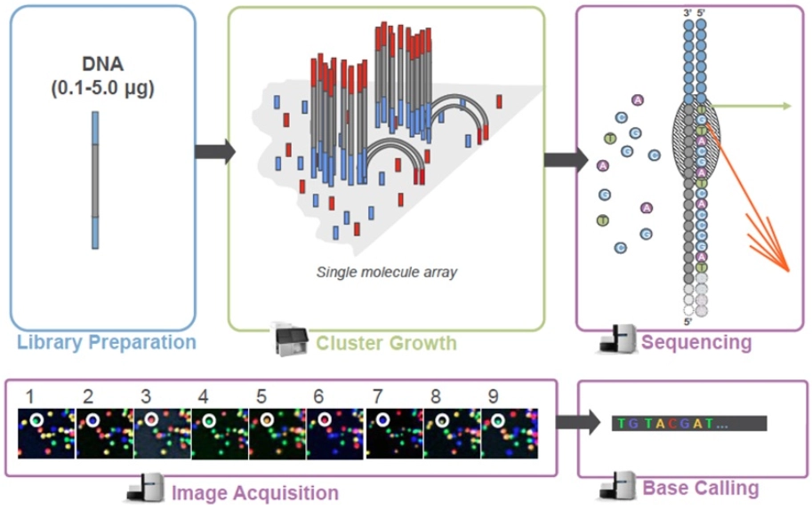
- Sequencing: The prepared library undergoes high-throughput sequencing using advanced platforms such as Illumina NovaSeq. Sequencing generates raw data on the chromatin accessibility landscape, providing a comprehensive view of open regions across the genome.
- Bioinformatics Analysis: The final step involves analyzing the sequencing data to interpret chromatin accessibility. This analysis includes several sub-steps to ensure accurate and meaningful results.

Fig 1. Sequencing process (source: https://www.illumina.com/)
Bioinformatics Analysis in ATAC-seq
Analyzing ATAC-seq data involves several important steps:
- Quality Control and Preprocessing: The initial step in bioinformatics analysis is to assess the quality of the sequencing reads. Tools like FastQC help identify issues such as low-quality bases or adapter contamination. Preprocessing steps, including trimming and filtering, ensure that only high-quality reads are used for subsequent analysis.
- Alignment: Cleaned reads are aligned to a reference genome using alignment tools such as Bowtie2 or BWA. Accurate alignment is essential for mapping reads to specific genomic locations and identifying accessible regions.
- Peak Calling: Peak calling identifies regions with significant read enrichment, which indicates open chromatin. Software such as MACS2 is used to differentiate true signals from background noise, pinpointing accessible regions accurately.
- Data Normalization and Comparison: To ensure that observed differences in chromatin accessibility are biologically meaningful, data normalization adjusts for technical variations such as differences in sequencing depth. Methods like read count normalization or RPM (Reads Per Million) are employed.
- Annotation and Visualization: Identified peaks are annotated with respect to genomic features such as genes, promoters, and transcription factor binding sites. Tools like HOMER assist in this process, while visualization tools like Integrative Genomics Viewer (IGV) or UCSC Genome Browser help researchers explore and interpret the data.
- Functional Enrichment Analysis: This step involves analyzing the biological significance of the accessible regions. By identifying enriched pathways, gene ontology terms, or transcription factor binding motifs, researchers can gain insights into the functional roles of these regions. Tools like GOseq or KOBAS are used for this purpose.
- Integration with Other Data Types: Integrating ATAC-seq data with other omics datasets, such as RNA-seq or ChIP-seq, provides a more comprehensive view of cellular processes. For instance, combining ATAC-seq with RNA-seq data can correlate chromatin accessibility with gene expression, while integrating it with ChIP-seq data can reveal interactions between transcription factors and chromatin modifications.
Applications of ATAC-seq
ATAC-seq is a versatile technique with a wide range of applications in epigenetic research. It provides valuable insights into nucleosome positioning and chromatin structure, which are crucial for understanding gene regulation and transcriptional control. By identifying key regulatory elements such as enhancers, promoters, and transcription factor binding sites, ATAC-seq helps in studying gene regulation and identifying potential therapeutic targets.
In a recently published study, ATAC-seq was employed to analyze chromatin accessibility in human embryonic stem cells and during their differentiation into definitive endoderm. The authors discovered that chromatin accessibility changes correlated with differentiation, revealing which genomic regions became accessible. It was found that nearly all non-coding RNAs (ncRNAs) interact with nearby genes, indicating a localized regulatory role rather than widespread effects. Additionally, the study identified thousands of unannotated RNAs that dynamically interact with chromatin, suggesting complex regulatory mechanisms that challenge simpler models of gene regulation, where a single ncRNA directly influences gene expression.[1]
In disease research, ATAC-seq can reveal changes in chromatin accessibility associated with conditions such as cancer. Identifying accessible regions in tumor cells can offer insights into cancer-specific regulatory mechanisms and potential biomarkers. [2] Additionally, ATAC-seq is useful for understanding how chromatin accessibility changes during development or in response to environmental stimuli, shedding light on mechanisms of cellular differentiation and adaptation.
Advantages of ATAC-seq
ATAC-seq offers several significant advantages over traditional methods. It provides high sensitivity in detecting chromatin accessibility, even in small cell populations. The technique requires only a minimal number of cells, making it suitable for studies involving rare or difficult-to-obtain samples. The entire workflow, from sample preparation to data analysis, can be completed in approximately three hours, making ATAC-seq a rapid and efficient method. Furthermore, ATAC-seq delivers comprehensive data that offers a detailed view of the chromatin landscape, contributing to a deeper understanding of gene regulation and epigenetic control.
Challenges and Considerations
Despite its strengths, ATAC-seq is not without challenges. Ensuring high-quality samples is crucial for obtaining reliable results; poor sample preparation can lead to artifacts or low-quality data. Additionally, the complexity of chromatin accessibility data requires careful analysis and interpretation. Integrating ATAC-seq data with other datasets can enhance insights but also adds to the analytical complexity.
Conclusion
ATAC-seq stands out as an innovative and efficient technique for exploring chromatin accessibility and understanding the regulatory landscape of the genome. Its ability to provide detailed insights into gene regulation, cellular differentiation, and disease mechanisms makes it an invaluable tool for researchers in genomics and molecular biology. By offering a comprehensive view of chromatin structure and function, ATAC-seq contributes significantly to advancing our knowledge of gene regulation and epigenetic control.
References
- Limouse, C., Smith, O. K., Jukam, D., Fryer, K. A., Greenleaf, W. J., & Straight, A. F. (2023). Global mapping of RNA-chromatin contacts reveals a proximity-dominated connectivity model for ncRNA-gene interactions. Nature communications, 14(1), 6073. https://doi.org/10.1038/s41467-023-41848-9
- Corces, M. R., Granja, J. M., Shams, S., Louie, B. H., Seoane, J. A., Zhou, W., Silva, T. C., Groeneveld, C., Wong, C. K., Cho, S. W., Satpathy, A. T., Mumbach, M. R., Hoadley, K. A., Robertson, A. G., Sheffield, N. C., Felau, I., Castro, M. a. A., Berman, B. P., Staudt, L. M., . . . Zhu, J. (2018). The chromatin accessibility landscape of primary human cancers. Science, 362(6413). https://doi.org/10.1126/science.aav1898
